-
News & Updates
News
May 30, 2018
June is Scoliosis Awareness Month

June is Scoliosis Awareness month; here is what you can do.
Please join us in bringing awareness to those with scoliosis that surgery is not the only option. Despite multiple studies have shown that over a 30 year period, scoliosis surgery adversely affected the quality of life scores when compared to those that chose to live well with scoliosis. The orthopedic insistence that surgery is the only option has handcuffed the families and patients into accepting a treatment they knew wasn’t the best option for them. Today things are changing. Thanks to our patients who spoke up and let people know of the their success. Use your personal social media platform this month to make people aware that scoliosis can be managed without surgery. Here are 3 things you can do to help.
Things to do during Scoliosis Awareness month (June)
-
Post a picture of yourself doing something inspiring and tell your story
-
Write a testimonial and send it to us and we’ll post your story
-
Make a donation to the non profit Scoliosis care Foundation
Together we can help people help themselves. Thank you for your involvement.
Drs Marc Lamantia, Gary Deutchman, Brett Diaz, Daniel Pagan, Arielle Klig, Kelsey Wiginton, Gigi Leonte, Steven Geanopulos, Morgan Maddox
December 1, 2014
What’s so good about the Nu-Schroth Scoliosis Method?
I recently took a call from a parent of an 11 year old diagnosed with scoliosis when she was 7 years old. During the past 4 years she has been under the care of an orthopedist who has dutifully monitored the child with x-rays during that time. At the last meeting the doctor recommended the child try some Schroth scoliosis exercises and recommended they find a local therapist trained in the 94 year old method. What changed? why did the Orthopedist suddenly recommend a nearly 100 year old method after 4 years of watchful waiting. This type of phone call has become quite common since the 2014 New York Times Article highlighting the Schroth scoliosis Method. The article is responsible for this resurgence in interest in non-surgical scoliosis treatment. In true American Fashion, we are not trying to extract out the “nutrient from the nutritious.” Let me explain, the Schroth method is an immersion technique and is provided in an in-patient setting. It includes up to 6 hours a day of therapy, massage, exercise and chiropractic. I spent two weeks at the clinic in 2008 and learned the method well. I knew when returning home I couldn’t offer an in-patient service and that I needed to modify the Schroth method to be suitable for U.S. consumption, and I didn’t want to water it down the way I knew would eventually happen. That’s when I came up with the “Nu-Schroth Method.”
The nu-schroth method uses the concepts I learned from the original Schroth method, but the concepts are applied with a 21st century take on rehabilitation. Lets face it, we’ve learned a tremendous amount since 1920, especially in the field or rehabilitation and the brain. The Nu-Schroth method uses contemporary rehabilitation methods like the Bosu ball, TRX training straps, HRV monitoring and vestibular rehabilitation. We teach the method in groups just the way Katarina Schroth did, and we modify activities of daily living the way Katarina Schroth did.
Here are 5 things that make the Nu-Schroth method the best even better than the Original Schroth Method for scoliosis;
1. The Nu-Schroth scoliosis method is taught so patients and families can do the exercises at home without needing to visit a therapists office daily. Exercises are broken down into Neurologic and Orthograde. Gait analysis is used as biofeedback to re-educate the gait of persons individually.
2. The Nu-Schroth scoliosis method uses Raster Stereography (a harmless imaging system) to monitor postural change, reducing the amount of harmful radiation exposure.
3. Nu-Schroth scoliosis method includes Chiropractic Neurology and manual therapy techniques which have been shown to improve the efficacy of exercises and postural restoration.
4. Nu-Schroth scoliosis method uses HRV monitoring to determine when patients should be resting, over training is a major cause of scoliosis progression
5. Nu-Schroth scoliosis method does their exercises to fatigue, then layers on complications to maximize results.
To learn more contact me directly at 1-800-281-5010 or visit scoliosissystems.com. 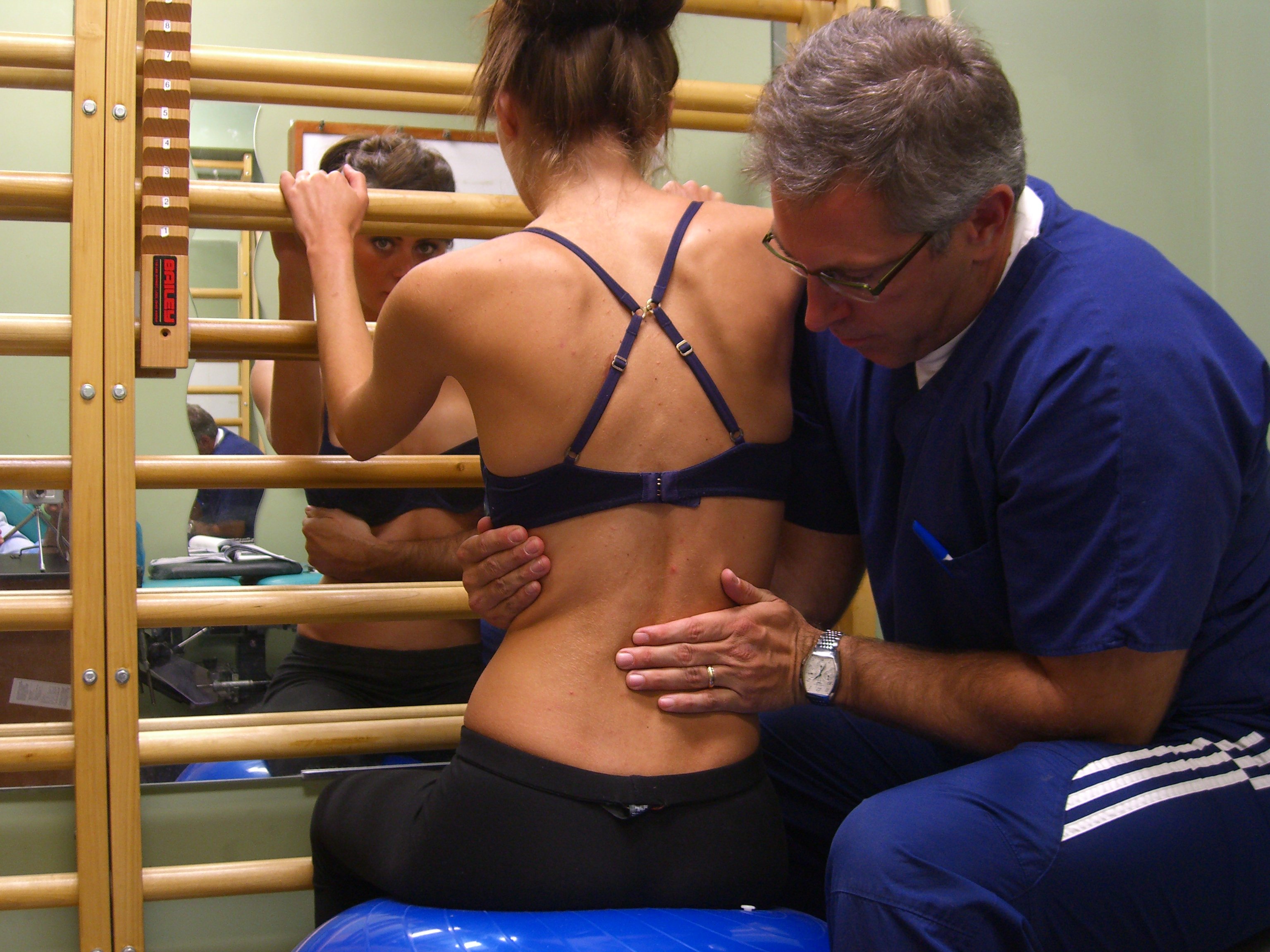
February 28, 2014
MRI For Scoliosis; Safe and Accurate
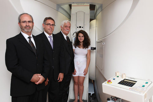
In photo: Dr. Gary Deutchman, Dr. Marc Lamantia, Dr. Raymond Damadian with patient.
MRI is not new, however Fonar Inc. has recently released their Scoliosis Coil, a modification which makes MRI useful in the measurement of Cobb Angles.
February 28, 2014
Clinical trials at a number of institutions in the United States
I would like to introduce you to information on our new scoliosis coil and screening/follow-up imaging application.
The scoliosis application uses a multi-channel, phased array, 80 cm, planar RF coil. This coil allows imaging of the entire spine (c-1 to s-1) in the standing upright position or seated upright position.
The scoliosis application allows for screening and follow-up of the patient without exposure to ionizing radiation. The entire study can be completed in 10 minutes or less resulting in economic feasibility for MRI. The multi planar MRI results provide additional information, not available from current P-A and lateral flat plane x-rays.
with severe cases of spinal abnormalities. Fonar welcomes any suggestions or insight that would aid in the development of spinal abnormalities applications.
February 28, 2014
FONAR Invents Radiation-Free Method to Diagnose and Monitor Scoliosis
Breakthrough Can End Later-Life Breast Cancer Induced by Overexposure To Spine X-Rays
MELVILLE, NEW YORK, November 26, 2007 – FONAR Corporation (NASDAQ-FONR), The Inventor of MR Scanning™, announced today at the 93rd meeting of the Radiological Society of North America (RSNA) in Chicago a groundbreaking invention in the diagnosis and monitoring of scoliosis. The patent-pending breakthrough utilizes new software and a new receiver coil developed for the unique FONAR UPRIGHT® Multi-Position™ MRI. The dramatic result is a single picture of the entire spine in the upright position (SEE PHOTO).
 |
|
Upright Scan of Scoliosis Patient with the Fonar Dynamic™ Upright® MRI
|
A National Cancer Institute (NCI) Report shows a 70% higher risk of breast cancer for women with scoliosis (www.cancer.gov/newscenter/scoliosis2000). The NCI report says, “Researchers have found that women with scoliosis, or abnormal curvature of the spine, who were exposed to multiple diagnostic X-rays during childhood and adolescence may be at increased risk of dying of breast cancer…. The 5,466 women in the study, who received an average of 24.7 X-rays, were found to have a 70% higher risk of breast cancer than women in the general population.” The report goes on to say that “although radiation exposures to breast tissue are much lower today than during the time period covered by this study, they are not insignificant.”
Current medical practice consists of ordering baseline X-rays for suspicious physical findings in children. (http://jaapa.com/issues/j20030901/articles/scoliosis.html). With X-ray, a scoliosis patient has a PA (posterior-anterior) and lateral radiograph two or three times a year. To reduce exposure to radiation, the patient is usually scanned with her back to the source of the X-ray.
Scoliosis affects 2-3% of the population or an estimated 6 million people in the United States, according to the Scoliosis Care Foundation (http://www.scoliosiscare.org/)
Raymond Damadian, M.D., president and founder of FONAR, said, “I’m delighted to announce our invention for the radiation-free evaluation of scoliosis with the FONAR Dynamic™ UPRIGHT® MRI. An accurate evaluation of scoliosis requires the patient to be upright. A conventional recumbent-only static MRI cannot meet this need. Of critical importance, our radiation-free application can be performed in the same amount of time and at the same cost as diagnosis and monitoring by X-ray. I believe it’s imperative that every hospital and practice performing scoliosis examinations consider providing their patients with the radiation-free choice that is finally available because of the unique benefits of the FONAR Dynamic™ UPRIGHT® MRI.”
The FONAR images provide coronal, sagittal and axial views of the entire spine – with no radiation. The 3-plane visualizations are achieved by 3-D acquisition with curved multi-planar reconstruction. Both the Cobb angles and the angular rotation of the vertebrae are measured. The FONAR UPRIGHT® MRI has another important advantage over X-ray. It sees, not only the curvature of the vertebrae, but the soft tissue, including the spinal cord, intervertebral discs, nerve roots and spinal ligaments.
“This application to scoliosis is just the latest of many unique advantages found only on the FONAR UPRIGHT® MRI,” continued Dr. Damadian. “Last week we reported on the landmark independent study by the UCLA School of Medicine, which reported the comparison of Dynamic™ UPRIGHT® MRI with static Upright MRI in more than 1,000 patients (1,301). A significant overall ‘miss rate’ of 18.1% by static MRI was cited. This large study proves the diagnostic advantages of FONAR UPRIGHT® Multi-Position™ weight-bearing MR imaging.”
FONAR plans to advertise its groundbreaking radiation-free scoliosis application in major medical magazines. To see the advertisement visit: http://www.fonar.com/news/pdf/scoliosis_ad.pdf.
As many as 60,000 people attend the Annual Meeting of The Radiological Society of North America. FONAR can be visited at booth 7753 in the South Hall at McCormick Place, Chicago, Illinois.