-
News & Updates
Vestibular
May 25, 2017
Scoliosis, Osteoporosis and Vertigo in Clinical Practice
Scoliosis, Osteoporosis and Vertigo in Clinical Practice
Do you have one or more of the following; Scoliosis, Osteoporosis or Vertigo? Clinicians and Researchers have long reported that all three of these conditions can occur simultaneously. And now, new research may answer at least in part why patients with Scoliosis also have Osteoporosis and Vertigo.
February 28, 2014
Improve the central neurological controls of posture
Vestibular problems resulting in; vertigo, dyslexia, balance disorders and postural disorganization, can respond well to treatment. Dr. Marc Lamantia is a Board Certified Chiropractic Neurologist with experience in the diagnosis and treatment of dizziness and other balance disorders. Our facilities are equipped with Video Infrared Electronystagmography (VNG); sophisticated diagnostic equipment to evaluate vestibular imbalances. Dr. Lamantia is an expert in the evaluation of vestibular testing, and will customize a treatment program based directly on your vestibular testing results. This is unique and often can be the difference between effective and ineffective treatment. Contact our offices to discuss your case. Below are so terms you may not be familiar with;
Vestibulo-Ocular Dysfunction: Research has linked vestibular and oculomotor dysfunction with dizziness, dyslexia, anxiety and Scoliosis. Hypoplasia of the brainstem may be an underlying cause when both scoliosis and ocular gaze palsy is present. MRI can be helpful in this diagnosis.
Vestibulo-Spinal Dysfunction: The Vestibular influence on lumbar spine and lower limb muscles have been found to be asymmetrical in patients with scoliosis and balance disorders. Appropriate evaluation can lead to functional rehabilitation and resolution of your problem.
Vestibular and Oculomotor Testing: Video Electronystagmography is the gold standard for evaluating vestibular and oculomotor function. Studies show well planned vestibular rehabilitation can effectively reduce vestibulo-ocular and vestibulo-spinal dysfunction. Dr. Lamantia has performed thousands of these proceedures and is considered a leader in his profession.
Vestibular Rehabilitation for Patients with Scoliosis: Postural habits of head tilt and asymmetric activation of muscles during movement can exacerbate scoliosis, and interfere with non-surgical reduction of scoliosis. Normal vestibular function is necessary to re-educate muscle recruitment patterns. Vestibular rehabilitation can be prescribed following a complete neurological evaluation including VNG and other clinical testing.
February 28, 2014
Vestibular Rehabilitation
Etiology of Idiopathic Scoliosis: Current Trends in Research.
(Lowe et al 2000)
…A number of studies have shown an abnormal nystagmus
response to caloric testing in patients with idiopathic scoliosis, suggesting an oculovestibular abnormality. Herman et al.46 proposed that a dysfunction of the motor cortex that controls axial posture results from a sensory input deficiency concerning spatial orientation and that this effect probably results from central proprioceptive sources involving visual and vestibular function. Other reports have supported this concept. The clinical syndrome of symmetrical horizontal or lateral gaze palsy is associated with a high prevalence of scoliosis of the idiopathic type. The site of neurological abnormality is thought to be the paramedian pontine reticular formation, which links the preocular motor nuclei and the vestibular nuclei. It is reasonable to speculate that the site of neuropathy in idiopathic scoliosis could also be the paramedian pontine reticular formation.
Vestibular Function in Adolescent Idiopathic Scoliosis
Abstract from Scoliosis Research Society (SRS) 2003 Meeting
Matthew T. Provencher M.D., Derin Wester, Ph.D., Bruce Gillingham M.D.; Naval Medical Center- San Diego, CA. Orthopedic Research and Education Foundation- Resident Research Grant
Conclusion: A central vestibular deficit is present in scoliosis patients. Central vestibular function is worse with larger curves, and the dysfunction is opposite to the curve. Curves with location in the mid-thoracic region demonstrated less central deficit than low-thoracic and lumbar scoliosis curves. The data supports a central vestibular dysfunction in patients with scoliosis
Spontaneous nystagmus (SN) and positional nystagmus (PN) were found in 24 out of the 47 patients with single curvatures and in only one subject in the control group (P less than 0.001).
Significant differences were observed in the caloric response between right and left scoliotic patients (P less than 0.05). The right convex patients had a sensitivity dominance in the right labyrinth and the left convex patients in the left labyrinth (Acta Orthop Scand 1979 Dec;50(6 Pt 2):759-69 Sahlstrand T, Petruson B.)
Vestibular mechanisms involved in idiopathic scoliosis:
(Arch Ital Biol 2002 Jan;140(1):67-80 Manzoni D, Miele F.Dipartimento di Fisiologia e Biochimica, Universita di Pisa, Via S. Zeno 31, I-56127 Pisa, Italy)
…It appears, however, that, in children, a slight imbalance in the activity of vestibular complex of both sides escapes the neuronal mechanisms responsible for vestibular compensation and leads to the spinal curvature which characterizes Idiopathic Scoliosis.
…The recommendation was made that a neurological examination, including assessment of vestibular function, be incorporated into screening methods for scoliosis.
(Jensen GM, Wilson KB. Phys Ther 1979 Oct;59(10):1226-33)
…Significant differences were found between patients with right convex curves and those with left convex curves in the distribution of eye predominance and in labyrinthine sensitivity
(Spine 1980 Nov-Dec;5(6):512-8 Sahlstrand T.)
IS THERE A RELATIONSHIP BETWEEN THE RESULTS OF UNTERBERGER TEST AND CONVEXITY OF SCOLIOSIS MAJOR CURVE?
Romano Michele, Zaina Fabio
ISICO (Italian Scientific Spine Institute), Via Carlo Crivelli 20, 20122 Milan, Italy – michele. [email protected]
Objective: The Unterberger stepping test is normally used to identify vestibular dysfunction and not to detect central disorders of balance. However we already made a previous study where we found a significant statistical difference in a sample of 30 scoliotic patient compared with a healthy control group. Our aim was to study if there is a relationship between direction of rotation during the test performance and convexity of scoliosis major curve.
Study design: 59 patient with adolescent idiopathic scoliosis (range: 14-55° Cobb) performed an Unterberger test (50 steps on place with closed eyes) before physical therapy session. Patients were divided into two groups: single curves, 29 subjects with 11 left and 18 right curves; double curves, 30 patients.
Results: There was a statistically significant concordance between the side of the curve and patient displacement after test performance in the single curves group when compared with the double curves, even if not all patients performed in the same way. There was not a significant statistical difference among left and right curve behaviours. Conclusion: These results could be explained both with neuro-motorial changes primary or secondary to the pathology, and biomechanical ones due to vertebral displacements.
February 28, 2014
neuro
What is Brain Lateralization?
In a study published in SPINE 1995, researchers tested linguistic processing, a higher cortical function, and concluded children with scoliosis had an entire brain dominance lateralized to one side, unlike unaffected children in the control group. This study demonstrates the need for advanced neuro-diagnostic testing and clinical correlations which leads to meaningful neurologically based rehabilitation.
Adolescent Idiopathic Scoliosis and Cerebral Asymmetry. An Examination of the non-spinal perceptual systems. SPINE 1995 Aug 1;20(15):1685-91.
| Dr. Lamantia Speaks at SOSORT |
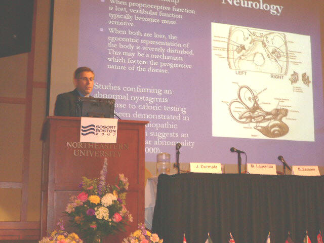
Dr. Lamantia presenting a report on the importance of vestibular rehabilitation at the 2007 International Conference on Conservative Management for Spinal Deformities at Northeastern University A comprehensive neurological examination is recommended although it is not mandatory to be fitted with an orthosis. Our approach to the non-surgical management of scoliosis is focused on neuro-muscular re-education of the postural support systems. The evaluation may include a sensory/motor exam, vestibular screening/evaluation, oculomotor screening/evaluation, movement analysis, neurocognitive asseessment, neurophysiological evaluations. Neurological patterns of dysfunction have been reported by researchers, however typical orthopedic management neglects most functional analyses and appropriate rehabilitation. In the event neurological imbalances are revealed, further testing, neuro-diagnostic imaging and appropriate therapy programs may be prescribed. |
February 28, 2014
Scoliosis Neurological Exams
Timothy C. Hain, MD. ![]() Page last modified: November 20, 2010
Page last modified: November 20, 2010
The Chiari I malformation, also known as the Arnold-Chiari malformation, is a relatively common syndrome caused by displacement of the cerebellar tonsils below the level of the foramen magnum. Associated with the Chiari malformation may be hydrocephalus, spina bifida, and syringomyelia. In most instances, symptoms present in middle age. There are hundreds of articles about this well known malformation in the literature (see Pubmed).
History and description:

In the early 1890s, Dr. Hans Chiari, professor of pathological anatomy at the German University in Prague, used autopsy specimens to describe four congenital anomalies later termed the Chiari malformations (types I to IV). Of these, the mildest (type -I) is the most common one encountered in clinical practice.
Nearly all Chiari malformations are type I. The type I Chiari malformation consists of caudal displacement of the cerebellar tonsils at least 3 mm into the upper cervical spinal canal . This type of herniation may be asymptomatic. The type II Chiari has a meningomyelocele (spinal opening). Type III has an encephalocele. (Ludwin and Norman, 1997)
CAUSE of the Chiari I malformation
The Chiari malformation is generally thought to be present from birth (although usually nobody knows this for sure). However, rarely mild Chiari malformations may result from low spinal fluid pressure. (Payner, 1994). It seems reasonable to us that Chiari’s may worsen gradually over life, and that a CSF leak might create a Chiari type of MRI picture as the brain droops down.
 |
 |
| Figure 1: Saggital midline MRI scan of person with Chiari-I malformation showing displacement of cerebellar tonsil below the upper margin of the foramen magnum | Normal MRI for comparison (obtained in 2009). Note alsothe improvement in MRI quality over 8 years. |
Rarely there is a hole in the brainstem/spinal cord associated with the Chiari malformation (see picture below). This is called a “syrinx”. A more subtle form of this is a “presyinx”, which is a potentially reversible state of spinal cord edema caused by obstruction to normal CSF flow pathways, especially in the cervical region (Goh et al, 2008).
 |
| Syrinx in cervical spinal cord of individual with an Arnold Chiari Malformation. Image courtesy of Ruth Ramsey, M.D. |
A closely related condition, basilar invagination may also be congenital or acquired (from arthritis).
DIAGNOSIS OF THE CHIARI MALFORMATION
Often when a person presents to the office with an undiagnosed Chiari, they have dizziness and headaches. This symptom complex triggers off a set of tests for inner ear conditions. During the clinical examination or perhaps on the ENG test, downbeating nystagmus is noticed at some point, and an MRI is obtained (see above). The MRI establishes the diagnosis.
 |
| Weak downbeating nystagmus seen in primary position during ENG, in patient with a prominent Chiari Malformation. In this patient the findings on clinical examination were much more obvious than this minimal DBN seen on ENG. |
 |
| Much stronger downbeating nystagmus seen on eccentric gaze, in patient with a prominent Chiari Malformation. There is no rebound nystagmus — he has a right-beating nystagmus that remains direction fixed on centering. |
The definitive method of diagnosis is with a T1 MRI scan of the posterior fossa, which documents the typical downward herniation of the cerebellar tonsils. A displacement of greater than 5 mm below the foramen magnum is considered significant. Occasionally Chiari symptoms occur in persons with lessened displacement of the tonsils (Milhorat et al, 1999). We are very unenthusiastic about surgical intervention in these patients (see later comments about the difference in enthusiasm between surgeons and others).
Curiously, the author has encountered patients from time to time in which the reading radiologist did not notice that the person had a Chiari malformation. In once case, it was a very substantial one. Because of this, we think it is prudent for the doctors who notice downbeating nystagmus on examination, to review the images of their patient’s MRI’s.
Symptoms of Chiari
Symptoms suggestive of Chiari include posterior headaches, dizziness and ataxia (especially associated with straining), fainting with a cough, and weakness or numbness. A recent review of the otologic manifestations of Chiari in 16 patients indicated that 81% reported episodic aural fullness, 81% tinnitus, 69% vertigo, and 56% flutuating hearing. Headache was also common (about 80%).
![]() Movie of downbeating nystagmus (5 meg). This patient does not have a Chiari but has nystagmus that is typical for Chiari.
Movie of downbeating nystagmus (5 meg). This patient does not have a Chiari but has nystagmus that is typical for Chiari.
Some patients with Chiari develop symptoms (dizziness mainly) on straining. This pressure sensitivity symptom is also shared by persons with superior canal dehiscence (SCD). Oddly, a recent report suggests that the Chiari Malformation is far more common in SCD (Kuhn and Clenney, 2010) than the normal population. We think that this report is likely due to sampling bias (i.e. this isn’t generally true). Nevertheless, we think it it is prudent to look for Chiari in SCD with an MRI.
Signs of Chiari
Signs of a significant Chiari malformation often include downbeat nystagmus (see above), poor pursuit for age, and alternating skew deviation.
 |
| Poor and extremely asymmetrical horizontal pursuit in person with prominent Chiari. |
 |
| Poor vertical pursuit in person with prominent Chiari. Vertical pursuit is often poor in persons with no neurological disorder so the value of this is limited. |
Rebound nystagmus can often be seen with a video-ENG device where the eyes can be vewed in complete darkness.
![]() Movie of rebound nystagmus (43 meg).
Movie of rebound nystagmus (43 meg).
Occasionally Chiari patients will have sensorineural hearing loss (Hendrix, 1992). Positional nystagmusis common. It is most commonly downbeating or lateral beating.
Persons with Chiari may develop vertigo after spending some time with their head inclined on their trunk. Thus the Chiari can cause cervical vertigo.
In the Dix-Hallpike maneuver for BPPV, this may be recognized by seeing a delayed onset positional nystagmus. This nystagmus can also be looked for more specifically with the “vertebral artery test” — a misnomer in this particular situation.
Differential Diagnosis of downbeating nystagmus
Other causes of constant downbeating nystagmus that should generally be considered are migraine,paraneoplastic cerebellar degeneration, and anterior canal BPPV. Generally, the paraneoplastic differential is the most concerning as the nystagmus can be very strong and the consequences of missing a tumor high. Anterior canal BPPV nystagmus is not a major differential as it is present only lying flat (unlike Chiari). Migraine nystagmus is almost always weak although it is accompanied by headache like Chiari.
Chiari symptoms overlap with those of Meniere’s disease as well as migraine (Sperling et al, 2001)
RISK OF THE CHIARI
A very few individuals with the Chiari malformation develop progresive neurological symptoms. This is most commonly due to an enlarging syrinx — a hole in the spinal cord (see above). In our otoneurology practice in Chicago, it is extremely rare for us to refer patients for surgery. Rather, we generally make arrangements to follow people on a yearly basis.
In our opinion, lumbar punctures, epidural blocks and related procedures that might cause a spinal fluid leak should be avoided whenever practical in persons with known Chiari malformation. The reason to avoid these procedures is that they may worsen the Chiari.
TREATMENT OF CHIARI MALFORMATION
The literature concerning treatment is large and is almost completely the work of neurosurgeons. There have been several recent review articles concerning indications for surgical treatment (Haroun, Guarnieri et al. 2000, Tubbs and Oakes 2004). There are very few articles concerning conservative management.
The following are the author’s opinions:
- Surgery
- Decisions regarding surgery are best based on patient cost/benefit.
- The cost of brain surgery is substantial – medical risk, monetary cost, and time cost.
- The main benefit to Chiari surgery is prevention of progression of a condition that is rarely progressive.
- Chiari surgery is nearly always best avoided in persons who have no neurological signs referable to the posterior fossa. In other words, don’t operate for symptoms of headache or dizziness — only operate for progressive physical signs that are unequivocally due to the Chiari.
- Decisions regarding whether or not surgery is indicated are best made by non-surgeons (i.e. neurologists), as surgeons are often biased towards surgery due to their training, inclination towards active treatments, and other factors. It is safest to get a second opinion from a non-surgeon, unaffiliated with the surgeon. Ideally, ask a neurologist in a different city. An unbiased second opinion could avoid a lifetime of neurological injury.
- Decisions regarding surgery are best based on patient cost/benefit.
- Non-surgical treatment
- There is no medication that treats the underlying cause of the Chiari malformation (brain displacement downward). There are some medications for neck pain, headache and dizziness that may help with the symptoms.
- Physical therapy and chiropractic manipulation of the neck does not help the Chiari malformation. In fact, it may make matters worse. We have no objection to massage.
- Avoidance of activities that precipitates symptoms (such as straining, athletic activity requiring straining or involving forceful movements of the head on shoulders) is often useful. As examples, we would suggest that persons with Chiari malformations not lift heavy weights, or play football.
Who should take care of Chiari malformation patients ?
In our opinion, the Chiari malformation is a condition that should be monitored on a once/year basis by a neurologist, and best of all, by a specialized neurologist — an otoneurologist. The reason for this is that while a neurosurgeon may ultimately operate on a patient with a Chiari, the huge majority of patients do not need brain surgery for the Chiari. We are generally unenthusiastic about asking neurosurgeons to make treatment decisions about elective brain surgery, as of course, neurosurgeons are paid to do surgery. We think it is generally better to have non-surgeons decide whether elective surgery should be paid for by insurance companies.
We are familiar with the Chiari malformation in our clinical otoneurology practice.