-
Scoliosis Treatment Success
February 9, 2014
February 9, 2014
Scoliosis Treatment Success
Presentation: A 62 year old female presented herself without referral for the examination. Her medical history included multiple recent falls, adult progressive scoliosis and a possible diagnosis of Multiple Sclerosis from her medical neurologist.
Observations posture reveal chronic towing of the spine to the right, shuffling gate with wide stance and the need to constantly hold on for stability.
Exams: a comprehensive neurological exam was performed including a sensory motor exam, a cerebellar and brainstem cranial nerve exam with appropriate testing of vestibular and oculomotor function. Clinical findings were nomometric in regards the motor examination. Ranges of motion were restricted in an age appropriate manor and sensory exam was further unremarkable. Vidoe Electronystagmography allowed for the evaluation of the vestiublar Cranial nerve portion, and as well as oculomotor testing. Upon vision denial testing in the upright seated posiition the patient presented with a non fatiguing right beating nystagmus with not tornsional or vertical component. The nystagmus was not visible without vison denial goggles. Oculomotor testing revealed findings of an adduction lag in the left eye during optokinetic stimulus. Xrays of the spine revealed an atypical towering scoliosis with severe lumbar disc degeneration and hyperostosis of the weight bearing joints.
Chiropractic evaluation included stereo views of the full spine posture and radiological views of along the X,Z, and the Yz axes and were reviewed in clincial context to recent MRIs provided by the patient. An occipito-atlanto-axial misalignment was measured. Reflexive retraction of the lower limb in non weight bearing supine posture was observed, and nerve root swelling with associated allodynia was localized by palpation at the c1 nerve root region. furhtermore, nucheal rigidity on the ipsilateral posterior neck musculature ws observed.
Differential Diagnosis: Cerviical Subluxation complex, Adult progressive scoliosis and associated osteoarthritis, left unilateral vestibulopathy, demeyelenating of the medial longitudinal fasicular tract from the right adbucens nucleus to the left oculomotor nucleus. CSF flow disruption.
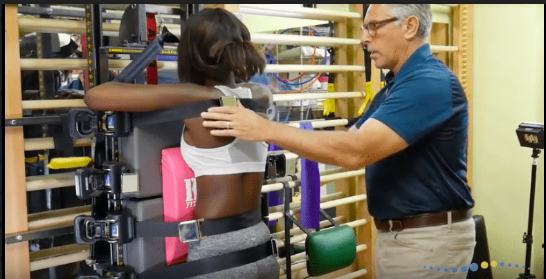
Plan: Follow up clincial testing is necessary. Xrays, caloric testing as well as repeat oculomotor testing is necessary to reproduce earlier findings. Chiropractic intervetion can begin immediately to reduce upper cervical spine dysfunction and C1 nerve root irritation. The patient is recommended to wear a spinal orthosis designed to improve dynamic control of the trunk and spine. Conservative treatment for vestibular and oculomotor findings.
0 Comments
Leave A Comment